Opinion Article - (2023) Volume 10, Issue 1
Received: 24-Feb-2023, Manuscript No. AJIROA-23-93289; Editor assigned: 27-Feb-2023, Pre QC No. AJIROA-23-93289 (PQ); Reviewed: 14-Mar-2023, QC No. AJIROA-23-93289; Revised: 21-Mar-2023, Manuscript No. AJIROA-23-93289 (R); Published: 28-Mar-2023
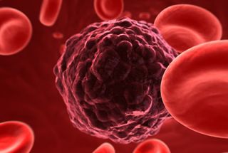
The development of a significant number of immature lymphocytes is one of the hallmarks of Acute lymphoblastic leukaemia (ALL), a blood cancer of the lymphoid line of blood cells. Besides from fatigue, symptoms may include pale skin, fever, easy bruising or bleeding, swollen lymph nodes, and bone discomfort. ALL develops quickly as an acute leukaemia and, if ignored, usually results in death within a few weeks or months. The cause is typically unknown. Down syndrome, Li-Fraumeni syndrome, and neurofibromatosis type 1 are examples of genetic risk factors. Risk factors related to the environment can include extensive radiation exposure or previous treatment. There is conflicting evidence on electromagnetic fields and pesticides. Some believe that a trigger could be an aberrant immunological response to a common virus. Several genetic alterations have a role in the underlying mechanism, which causes fast cell division. The bone marrow's overabundance of immature lymphocytes inhibits the growth of new red blood cells, white blood cells, and platelets. Bone marrow analysis and blood testing are frequently used to make a diagnosis. Chemotherapy is frequently used as the first treatment for ALL with the goal of achieving remission. Further chemotherapy often follows this over the course of several years. As systemic chemotherapy can only penetrate a limited amount of the central nervous system, and the central nervous system is a frequent site for acute lymphoblastic leukaemia relapse, intrathecal chemotherapy is typically used in addition to other forms of treatment.
Treatment
A lasting remission, which is defined as the absence of cancerous cells in the body (typically less than 5% blast cells in the bone marrow), is the goal of treatment.The effectiveness of treatment regimens has been improved over the past few decades, which has raised survival rates. Chemotherapy, steroids, radiation therapy, rigorous combination therapies (including bone marrow or stem cell transplants), targeted therapy, and/or growth hormones are a few possible treatments for acute leukaemia.
Diagnosis
A detailed medical history, physical examination, a full blood count, and blood smears are the first steps in making the diagnosis of ALL. While many of the symptoms of ALL are similar to those of common illnesses, persistent or unexplained symptoms should raise cancer suspicion.
Further testing is frequently required because many aspects of the medical history and exam that are not specific to ALL are present. Because they signify a quick development of lymphoid cells in the bone marrow, an abundance of white blood cells and lymphoblasts in the blood can be suspicious for ALL.
A worse prognosis is often indicated by numbers that are higher. Although white blood cell counts at the time of initial presentation can vary greatly, circulating lymphoblast cells are typically seen on peripheral blood smears.
The diagnosis of ALL is confirmed by a bone marrow biopsy, which often reveals >20% leukemic lymphoblasts among all cells. If the spinal column and/or brain have been invaded, it can be determined with a lumbar puncture, sometimes referred to as a spinal tap. Leukemic cells in the lumbar puncture or the previously mentioned clinical symptoms of central nervous system can both be used to confirm the involvement of the brain and spinal column. Blood count, renal function, electrolyte, and liver enzyme testing are just a few of the laboratory tests that could reveal anomalies.
If Philadelphia chromosome is present, cytogenetic analysis, and immunophenotyping determine if the leukemic cells are lymphoblastic (neutrophils, eosinophils, basophils, myeloblastic, neutrophils, eosinophils, basophils, B lymphocytes or T lymphocytes). Cytogenetic analysis of bone marrow samples can be used to diagnose diseases and forecast how aggressively they will progress. There have been various mutations linked to either shorter or longer survival. Common acute lymphoblastic leukemia antigens may be present on the surface of leukemic cells, as shown by immunohistochemical analysis.
Select your language of interest to view the total content in your interested language